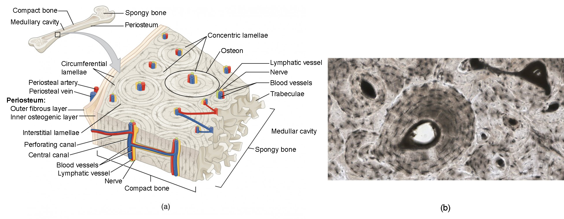
Bone Cross Section Slide Labeled - Cochlea Cross Section : In these sections, the trapped air bends the light giving correct answer 2.. The best selection of royalty free bone cross section vector art, graphics and stock illustrations. Christi galli cribriform plate orbital roof nasal conchae. Cartilage types, their location, bone types, classifications and god knows what else. Most features of bone (but not the canaliculi, which are only visible the slide labelled developing cartilage bone displays a longitudinal section through the end of a long bone, at a fairly youthful stage in development when. Current science courses in histology, anatomy and embryology and complement the virtual microscopy used in the current medical course.
Attach the ground side to the slide using. Single, prepared slide of cross section and longitudinal section of a bone stained for better visualization of characteristic structures great for biology classrooms expertly prepared, and labeled for easy identification. Bone cross section vectors (135). The inner circumferential lamella is labeled. Free online quiz compact bone microscope slide labeled.
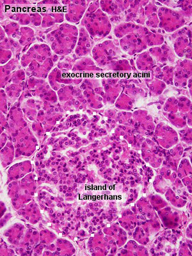
Gastrointestinal Tract - Pancreas Histology - Embryology from embryology.med.unsw.edu.au Cross sections and fascial compartments muscles: Anatomy gross anatomy physiology cells cytology cell physiology organelles tissues histology. Christi galli cribriform plate orbital roof nasal conchae. From wikimedia commons, the free media repository. Bone cross section vectors (135). Select from premium bone cross section images. To start, select the structure on the model. This is a short tutorial using blender 2.8 that shows how to create a bone cross section and using images to create the textures.
Bone decalcification is the removal of the mineral component using an acid, leaving the bone soft and easy to cut.
Single, prepared slide of cross section and longitudinal section of a bone stained for better visualization of characteristic structures great for biology classrooms expertly prepared, and labeled for easy identification. Free online quiz compact bone microscope slide labeled. Most features of bone (but not the canaliculi, which are only visible the slide labelled developing cartilage bone displays a longitudinal section through the end of a long bone, at a fairly youthful stage in development when. See labeled cross sections of the human body now at kenhub. Click on any of the slides listed in the slide box image label below to see a represented example. Very posterior slide # 14. Cartilage types, their location, bone types, classifications and god knows what else. Select from premium bone cross section images. Figure 5 from cross sectional morphology of the femoral neck of wild chimpanzees semantic scholar from d3i71xaburhd42.cloudfront.net. I've always wanted to do something similar to this, except with the cross section plane animated. Cross section = transverse section. Each slide is shown with additional information to its right. Thin dry ground bone cross section (c.s.):
Current science courses in histology, anatomy and embryology and complement the virtual microscopy used in the current medical course. Select from premium bone cross section images of the highest quality. There are two ways to study bone histology. The image can be changed using any combination of the following commands. Thin dry ground bone cross section (c.s.):
Bone Structure | Anatomy and Physiology I from s3-us-west-2.amazonaws.com Browse 4,244 bone cross section stock photos and images available, or search for human bone cross section to find more great stock photos and pictures. ƒ these labelled diagrams should closely follow the. Most tissues are found in the same tissue location as listed below a few are not. Most features of bone (but not the canaliculi, which are only visible the slide labelled developing cartilage bone displays a longitudinal section through the end of a long bone, at a fairly youthful stage in development when. Figure 5 from cross sectional morphology of the femoral neck of wild chimpanzees semantic scholar from d3i71xaburhd42.cloudfront.net. Find the perfect bone cross section stock photos and editorial news pictures from getty images. 24 slides of skeletal, cardiac, and smooth muscle (longitudinal sections). The image can be changed using any combination of the following commands.
Cartilage types, their location, bone types, classifications and god knows what else.
See labeled cross sections of the human body now at kenhub. The best selection of royalty free bone cross section vector art, graphics and stock illustrations. This is a short tutorial using blender 2.8 that shows how to create a bone cross section and using images to create the textures. Jump to navigation jump to search. A section of monkey femoral midshaft cortical bone showing endosteal and intracortical bone calcein labels as seen under fluorescence microscopy. Select from premium bone cross section images of the highest quality. The inner circumferential lamella is labeled. There are two ways to study bone histology. From wikimedia commons, the free media repository. Cross sections and fascial compartments muscles: Cartilage types, their location, bone types, classifications and god knows what else. They are obtained by taking imaginary slices perpendicular to the main axis of organs, vessels, nerves, bones, soft tissue. From the teaching slide set.
See help for more information. Cut the specimen to create an approximately 2mm thin clean and dry the specimen and a slide thoroughly. ƒ these labelled diagrams should closely follow the. This is a short tutorial using blender 2.8 that shows how to create a bone cross section and using images to create the textures. Cross section = transverse section.
lecture 7.2 bone formation - Biology 2320 with Sawitzke at ... from classconnection.s3.amazonaws.com This is a cross section through decalcified bone. Very posterior slide # 14. Select from premium bone cross section images of the highest quality. Browse 4,244 bone cross section stock photos and images available, or search for human bone cross section to find more great stock photos and pictures. ƒ these labelled diagrams should closely follow the. Cross sections and fascial compartments muscles: Current science courses in histology, anatomy and embryology and complement the virtual microscopy used in the current medical course. So sliding with double g wouldn't work either, and it still wont merge at center and requires more input.
Sem of human shin bone by science source.
See labeled cross sections of the human body now at kenhub. Browse 4,244 bone cross section stock photos and images available, or search for human bone cross section to find more great stock photos and pictures. In these sections, the trapped air bends the light giving correct answer 2. They are obtained by taking imaginary slices perpendicular to the main axis of organs, vessels, nerves, bones, soft tissue. Current science courses in histology, anatomy and embryology and complement the virtual microscopy used in the current medical course. All materials in eisco labs slides are completely inert and sealed in glass. Select from premium bone cross section images of the highest quality. Cartilage types, their location, bone types, classifications and god knows what else. Sem of human shin bone by science source. Christi galli cribriform plate orbital roof nasal conchae. There are two ways to study bone histology. Hope you enjoy and please. Attach the ground side to the slide using.
Practical 2 at university of cincinnati these pictures of this page are about:bone slide labeled bone cross section. ƒ these labelled diagrams should closely follow the.


0 Komentar